
What is Spinal Regenerative Medicine?
Introduction
Inflammatory and mechanical trauma cause degeneration and aging of the spine including the cartilage, bones and discs. Surgery is typically only helpful in the end stages of this process where the damage is permanent and irreversible. Regenerative medicine, or the use of biologically active injections, aims to intervene before the tissue damage is so severe as to require surgery to fix it. Regenerative medicine uses mesenchymal stem cells and platelet rich plasma to drive the natural healing process. While there is great promise in such therapies, the current medical literature is just starting to support to the use of regenerative products in the spine (1). Platelet rich plasma (PRP) is the most widely used agent in regenerative medicine and shows promise in treating low back pain caused by facet arthritis. Stem cells have also been used with success, however the literature is not as robust as for platelet rich plasma. In this post we explore both options and the science supporting their use.
What is regenerative medicine?
As your body ages certain cell types become inactive leading to the loss of the regenerative capacity of your body to heal injury. Regenerative medicine uses biologically active treatments to jump-start or supplement these healing pathways that may have become damaged or lost. Regenerative medicine is appealing to patients with spine disease as current treatments are often either ineffective or require invasive surgery to treat end-stage problems. Regenerative medicine hopes to intervene earlier in the disease process to keep the spine from becoming so damaged it must be surgically repaired.
The two most popular types of regenerative medicine approaches include the use of stem-cells and platelet rich plasma (PRP). Both contain growth factors that can activate your cells to proliferate and heal tissues. These treatments generally involve injecting concentrations of your blood or bone marrow back into your body in different areas to accelerate and improve the healing process.
Regenerative medicine has taken off on the promise of restoring joints back to their original state with just the tip of a needle. Although the potential for these therapies remains enormous, and they can be useful in the correct setting, here we will review the nitty gritty evidence and see what really works.
What is a Stem Cell?
Your body consists trillions of small structures known as cells which are the essential building blocks of life. Most cells are specialized (i.e liver cells or brain cells), to serve a specific purpose. Stem cells are a special group of cells that your body keeps in reserve that can differentiate (turn into) any cells of the body. Stem cells are powerful as they are theoretically capable of replacing damaged or destroyed tissue. They serve as a reserve population of cells which divide when needed to replenish specialized tissue cells. There are two main types of stem cells: embryonic and mesenchymal.
Embryonic stem cells are derived from the epiblast layer of the embryo and can differentiate into all cells of the three germ layers. They are only found in the very early stages of the embryo. Unlike other cell types, they do not express immune signaling major histocompatibility complex (MHC) molecules making them “immune privileged (2).” Embryonic stem cells are only available for medical use if they were stored by your parents.
Mesenchymal stem cells (MSCs) are found in adults and are multipotent, meaning they can only differentiate into the cell types of the mesenchymal lineage (bone, cartilage, muscle, fat). They are sourced from the aspirating a sample of bone marrow from the pelvis, so called bone marrow derived mesenchymal stem cells (BM-MSC), and the cells are concentrated by spinning them in a device called a centrifuge. The bone marrow is the most commonly used source of stem cells and is Food and Drug Administration (FDA) approved (3). They can also be obtained from fat, so called adipose derived mesenchymal stem cells (AD-MSC). Rarer sources of MSCs are the placenta, dental pulp, and amniotic fluid (4). The clinical effects of varying sources of stem cells sources remains a hotly debated topic (3).
There are several challenges with getting stem cells to repair tissues. One of the main issues with implanting stem cells is that they can die soon after implantation (5). Another problem is getting them to differentiate into the tissues of choice and incorporate into the host tissues. In theory, stems cells sound promising, however, the research to support their routine use is in its infancy. Let’s explore how stem cells are currently used in clinical practice.
Degenerative disc disease:
The intervertebral disc is a ring structure that consists of two parts: the annulus fibrosis which is a thick multilayer fibrous ring and a central gel matrix known as the nucleus pulposus. For the purposes of this paper we will focus on the gel matrix because it is critical for maintaining the integrity of the disc.
In a normal intervertebral disc, there is a population of special cells known as notochordal cells which are responsible for generating the gel matrix. The gel matrix is unique because it consists of molecules known as glycosaminoglycans (GAG) that keeps discs hydrated. Glycosaminoglycans (such as aggrecan) are directly responsible for the water content of the discs and hydrostatic pressure to ensure discs can help absorb the shocks of daily life. This area of the disc is notably avascular, meaning it does not have a good blood supply and thus is a low oxygen environment. Slight changes in pH (the acidity of the disc), or microtrauma can damage the sensitive nucleus pulposus cells, initiating degenerative changes that are like a one way avalanche.
For a variety of reasons, these cells can die off as you age which kicks starts the degenerative process because the discs lose the ability to produce GAGs. Without the gel matrix, discs cannot absorb water and they become dried out and shrivel. As the discs shrivel, they become mechanically unstable which can accelerate degeneration from even trivial forces of day to day living. In this scenario, inflammation continues to increase leading to further destruction.
How can stem cells be used clinically?
How can we use stem cells? Stem cells are potentially beneficial because they can become almost any tissue of the body. So why are we not using these all the time in routine treatment? The answer is that although there is promise in preclinical and clinical studies, stem cell therapy research is ongoing.
The idea to use stem cells comes from the concept that the nucleus pulposus cells are important for keeping an intervertebral disc viable. They generate the extracellular matrix that helps keep the discs hydrated. When they die off the disc enters a reinforcing degeneration downward spiral (6). So, the idea is to inject stem cells which can differentiate into nucleus pulposus cells and take the place of the ones that have died. The issue is how do you get stem cells to transform into nucleus pulposus cells and how do you get them to survive long enough to do the job. For this reason intradiscal injection of stem cells is a promising therapy that needs further development (3).
Stem cells seem to act not only by differentiation, but also through signaling—getting other nearby cells to do the work of tissue repair. They can do this through immunomodulation, trophic and anticatabolic effects. MSCS can stimulate nearby cells to generate more disc matrix and prevent damage from inflammation through immunomodulation (7). Therefore long term survival of the stem cells may not be clinically relevant.
Using a transcription factor known as growth factor-6 (GF-6) scientists were able to induce BM-MSC and AD-MSC into nucleus pulposus resembling cells (8). These cells have shown promise in animal models, where their injection has improved the radiographic appearance of discs in terms of improved disc height and hydration (9,10). However, are these results translatable to humans?
Pilot studies have been performed using BM-MSCs implanted into the human subjects with symptomatic degenerative disc disease showing that overall it was safe and reduced pain (11–13).
Following the pilot studies, randomized studies have been performed showing improved pain and functional outcomes in stem cell injection for patients with chronic low back pain and moderate degenerative disc disease (14,15). These studies are limited in that they had very small numbers of participants (only 24 patients) but do offer some evidence that stem cells can be beneficial. Meta-analyses of compiled data of stem cell injection intradiscally show improvement in pain scores up to 12 months (16).
Remaining questions in the field of stem cell regenerative medicine include how long stem cells survive once injected into biologically and mechanically hostile environment of the intervertebral disc. Canine studies have shown that stem cells only survive on average for 3 weeks (17). The environment in a degenerative disc is typically one of low oxygen, making survival of MSCs less likely. Preconditioning cells for a hypoxic environment may be a useful way to extend their lifespan in the body (18).
If Stem Cells Only Survive for a Short Period, How do They Produce Pain Relief?
It is thought that stem cells do not act by regenerating tissues but rather by secreting signaling molecules that reactivate nearby degenerated tissue. Injection of growth factors were previously thought to be a viable treatment for disc disease however the effects are so short lived that it’s not considered a useful clinical approach. Instead it is possible that the use of MSCS function as small growth factor factories that help adjacent tissues (4). MSCs do this by excreting paracrine factors and exomes which reduce inflammation, inhibit programmed cell death and stimulate cellular proliferation (4).
Exomes are small vesicles released from the cell that contain signaling molecules and nucleic acid (DNA and RNA). Exomes are then absorbed by nearby cells through endocytosis or direct plasma membrane fusion and can transfer genetic material. Exomes can inhibit cell inflammation and oxidative stress, while stimulating the production of cell matrix synthesis and encouraging cell division by transferring genetic material and signaling molecules to other weary cells (4). The use of stem cell exomes has emerged as an independent treatment for degenerative disc disease and shows promising results in animal models (4,19). Exomes can help maintain disc homeostasis by inhibiting apoptosis of nucleus pulposus cells through antioxidant and anti-inflammatory properties.
Clinical usage of exomes have several advantages over MSCs. Exomes are much smaller and thus easier to store and have less issues with cellular senescence (cell aging). Exomes have little issues with immunogenicity and are considered safer than MSCs because of no tumor forming potential (4). Exomes show promise in in vitro studies for cartilage repair when combined with hydrogels (20).
Despite the promise of exomes, they are not totally ready for clinical applications as of this writing. Heavily manipulated stem cells and products are currently not approved by the FDA (3). Challenges include the need for suitable donor MSCs which the appropriate biochemical profile to match the diverse pathogenesis of disc degeneration. Manufacturing, purification and standardization processes for exomes are still in their infancy. The appropriate dosing, toxicity, and routes of administration for exomes are also currently unknown.
What is Platelet Rich Plasma (PRP)?
Platelets (thrombocytes) are cell fragments that come from a large megakaryocyte cells and are involved in hemostasis, wound healing, and tissue regeneration. Platelets contain a dense amount of alpha granules (small packets of chemicals) that are released on activation. Platelets go through several stages: adhesion, aggregation, and activation. On activation they change their shape to release the granules (21). Platelets can interact with other cells including endothelial and immune cells and can also target bacteria to trigger an immune response via toll like receptors.
Platelet activation is a crucial first step in repairing an injury and is responsible for the release of growth factors such as platelet derived growth factor (PDGF) and transforming growth factor beta 1 (TGF-1). Growth factors lead to the activation of other cell populations such as stem cells and fibroblast which results in extracellular matrix formation such as new collagen and bone production (22,23).
TABLE OF PLATELET CONTENTS:
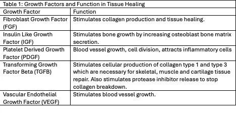
Platelet rich plasma is a fraction of blood containing concentration a high of platelets. A routine blood draw is performed and then processed in a machine called a centrifuge to allow a 2.5 times increase in platelet concentration (3). It is currently unknown the exact proportions of the various growth factors that are required to stimulate healing and these levels are different in everyone. Thus it is important to remember that your personal chemical milieu can impact the variability of efficacy and is influenced by things such as your age, gender, health, nutritional status (24).
Platelet rich plasma is safe and effective for the treatment of low back pain originating from lumbar facet arthritis, intervertebral disc disease, or sacroiliac joint dysfunction. It can also be used to treat sciatica and low back pain originating from epidural nerve compression (25).
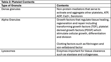
Platelet rich plasma is the most commonly used biologic to treat lumbar facet disease (arthritis affecting the small joints in the posterior aspect of the back), sacroiliac disease and seems to have longer lasting pain relief compared to steroids (26). Platelet rich plasma is commonly used to treat arthritic disorders and has been shown in a large metanalysis of 34 studies to improve knee osteoarthritis pain compared to a placebo (27). PRP injected into the disc itself has not been shown to improve pain in randomized controlled trials. A systematic review performed on PRP injected into the epidural space did show longer lasting pain relief compared to epidural steroid (29). Meta-analyses of PRP injected into the facet joints did show long term (12-month) improvement in low back pain compared to controls (16).
There are some factors that go into the quality of platelets that can be obtained from the body. Typically blood is obtained from a peripheral source such as a vein in the arm which has been shown to be the best site to obtain concentrated platelets (30). A large needle is used to prevent the platelets from breaking down when they are extracted. It should also be noted that the quality of your platelets is dependent on several factors such as your age, sex, medication use (such as anticoagulants, and anti-inflammatories), and not everyone will have the same number or quality of platelets (21). Prior to your treatment with PRP it may be necessary to stop certain antiplatelet drugs such as aspirin and clopidogrel.
Once the sample has been isolated (the amount drawn is dependent on the use and desired volume), the blood is combined with an anticoagulant (acid citrate dextrose). The blood is then introduced into a sterile centrifuge which spins the liquid at high speeds to separate the cellular layers. Red blood cells fall to the bottom, whereas platelets arrive at the top. Platelets may be chemically or physically activated prior to injection however this has not shown to be beneficial, and we thus use them in their native form (21).
The clinical effect of PRP may take up to one month to kick in (31).
Who should check with their doctor before having PRP or BMAC injections:
In general both PRP and BMAC injections are very safe, however there are some patients who should have a careful discussion with their physician before a regenerative injection is performed (32).
Patients with an active viral infection such as hepatitis or human immunodeficiency virus (HIV) may still have PRP injections if the virus has been treated and certain criteria are met. For HIV, the virus must be undetectable and the CD4 blood count must be greater than 350/mm3.
Patients with untreated active bacterial or viral injections should not have PRP injections performed. If you have active fever, chills, or other signs of injection please check with your physician before having a PRP injection.
If you are on dialysis or have kidney failure you must be carefully monitored for infection following a PRP injection.
Any patient with active cancer (solid tumor, blood cancer, or other malignancy) should check with their oncologist and physician before having a PRP injection.
Patients with bleeding or clotting disorders, or who are actively on blood thinners or antiplatelet medications should check with their physician before having PRP. All antiplatelet and anticoagulants should be held prior to having a PRP or BMAC injection.
The Safety of regenerative medicine
The rate of adverse events with platelet rich plasma has been reported to be as high as 6.3%, the most common issue being temporary post-procedural pain and inflammation. Because platelet rich plasma is a component of blood it is possible for it to contain inflammatory white blood cells which can accentuate the inflammatory process. Other complications can include muscle stiffness, numbness, positional headaches, and skin reactions. In one series of lumbar facet injections of PRP, no serious complications were noted including nerve injury, rejection reaction, or infection. The most common adverse event was temporary exacerbation of back pain. Because PRP and BMAC are totally autologous, meaning they comes from your own body, the potential for side effects compared to other common pain medications such as steroid cortisone is low (33,34).
Stem cell implantation also seems relatively safe with the most common adverse event within 1 year being a temporary increase in pain 3-5 days after the injection (35).
Some things to keep in mind if you are a patient:
1. Although they are at the forefront of regenerative medicine, remember that biologics and stem cell therapies are not miracle cures. It is important to have realistic expectations. They can be helpful for reducing pain but will not cure severely damaged areas of your back/neck.
2. Biologic therapy has variable effects depending on the health of your own cells. You may require more than one injection to treat the damaged area of your back for the therapy to be effective.
3. Please tell your doctor if you are on any antiplatelet or anticoagulation medications such as aspirin, warfarin, or heparin. These should be discontinued prior to your procedure to reduce the risks of bleeding.
4. It is important to remember that you should discontinue non-steroidal anti-inflammatory medications for 2 weeks after your procedure to ensure the effectiveness of the biologic. Anti-inflammatory medications will mitigate tissue healing. As such your pain may increase in the short term.
5. Other risks include damage to nerves and blood vessels near the injection area, infection, and bleeding.
6. Cancer is a theoretical concern for stem cell transplantation, however it has not been observed in studies after stem cell injection (3).
Conclusions
Regenerative medicine is an exciting new area of treatment for degenerative spinal conditions. Although current clinical data is promising, more studies focusing on how to harness its potential appropriately are needed. Overall, biologic injections appear relatively safe and have the possibility of improved pain and function.
References:
1. Maloney J, Strand N, Wie C, Pew S, Dawodu A, Dunn T, et al. Current Review of Regenerative Medicine Therapies for Spine-Related Pain. Curr Pain Headache Rep. 2024 Sep;28(9):949–55. doi:10.1007/s11916-023-01194-3
2. Fernández-Francos S, Eiro N, Costa LA, Escudero-Cernuda S, Fernández-Sánchez ML, Vizoso FJ. Mesenchymal Stem Cells as a Cornerstone in a Galaxy of Intercellular Signals: Basis for a New Era of Medicine. IJMS. 2021 Mar 30;22(7):3576. doi:10.3390/ijms22073576
3. Manchikanti L. Responsible, Safe, and Effective Use ofBiologics in the Management of Low BackPain: American Society of Interventional PainPhysicians (ASIPP) Guidelines. Pain Phys. 2019 Jan 11;22(22;1s):s1–74. doi:10.36076/ppj/2019.22.s1
4. Bhujel B, Shin HE, Choi DJ, Han I. Mesenchymal Stem Cell-Derived Exosomes and Intervertebral Disc Regeneration: Review. IJMS. 2022 Jun 30;23(13):7306. doi:10.3390/ijms23137306
5. Munda M, Velnar T. Stem cell therapy for degenerative disc disease: Bridging the gap between preclinical promise and clinical potential. Biomol Biomed. 2024 Mar 11;24(2):210–8. doi:10.17305/bb.2023.9518
6. Hohaus C, Ganey TM, Minkus Y, Meisel HJ. Cell transplantation in lumbar spine disc degeneration disease. Eur Spine J. 2008 Dec;17(S4):492–503. doi:10.1007/s00586-008-0750-6
7. Barakat AH, Elwell VA, Lam KS. Stem cell therapy in discogenic back pain. J Spine Surg. 2019 Dec;5(4):561–83. doi:10.21037/jss.2019.09.22
8. Clarke LE, McConnell JC, Sherratt MJ, Derby B, Richardson SM, Hoyland JA. Growth differentiation factor 6 and transforming growth factor-beta differentially mediate mesenchymal stem cell differentiation, composition, and micromechanical properties of nucleus pulposus constructs. Arthritis Res Ther. 2014 Mar 12;16(2):R67. doi:10.1186/ar4505
9. Sakai D, Mochida J, Iwashina T, Watanabe T, Nakai T, Ando K, et al. Differentiation of mesenchymal stem cells transplanted to a rabbit degenerative disc model: potential and limitations for stem cell therapy in disc regeneration. Spine (Phila Pa 1976). 2005 Nov 1;30(21):2379–87. doi:10.1097/01.brs.0000184365.28481.e3 PubMed PMID: 16261113.
10. Crevensten G, Walsh AJL, Ananthakrishnan D, Page P, Wahba GM, Lotz JC, et al. Intervertebral disc cell therapy for regeneration: mesenchymal stem cell implantation in rat intervertebral discs. Ann Biomed Eng. 2004 Mar;32(3):430–4. doi:10.1023/b:abme.0000017545.84833.7c PubMed PMID: 15095817.
11. Centeno C, Markle J, Dodson E, Stemper I, Williams CJ, Hyzy M, et al. Treatment of lumbar degenerative disc disease-associated radicular pain with culture-expanded autologous mesenchymal stem cells: a pilot study on safety and efficacy. J Transl Med. 2017 Dec;15(1):197. doi:10.1186/s12967-017-1300-y
12. Haines CM, Bhatt FR, Orosz LD, Yamout T, Namian S, Bharara N, et al. Low Back Pain, Disability, and Quality of Life One Year following Intradiscal Injection of Autologous Bone Marrow Aspirate Concentrate. Chapel A, editor. Stem Cells International. 2022 Dec 12;2022:1–7. doi:10.1155/2022/9617511
13. Wolff M, Shillington JM, Rathbone C, Piasecki SK, Barnes B. Injections of concentrated bone marrow aspirate as treatment for Discogenic pain: a retrospective analysis. BMC Musculoskelet Disord. 2020 Dec;21(1):135. doi:10.1186/s12891-020-3126-7
14. Noriega DC, Ardura F, Hernández-Ramajo R, Martín-Ferrero MÁ, Sánchez-Lite I, Toribio B, et al. Intervertebral Disc Repair by Allogeneic Mesenchymal Bone Marrow Cells: A Randomized Controlled Trial. Transplantation. 2017 Aug;101(8):1945–51. doi:10.1097/TP.0000000000001484 PubMed PMID: 27661661.
15. Amirdelfan K, Bae H, McJunkin T, DePalma M, Kim K, Beckworth WJ, et al. Allogeneic mesenchymal precursor cells treatment for chronic low back pain associated with degenerative disc disease: a prospective randomized, placebo-controlled 36-month study of safety and efficacy. Spine J. 2021 Feb;21(2):212–30. doi:10.1016/j.spinee.2020.10.004 PubMed PMID: 33045417.
16. Manchikanti L. Do Regenerative Medicine Therapies ProvideLong-Term Relief in Chronic Low Back Pain: ASystematic Review and Metaanalysis. Pain Phys. 2018 Nov 14;1(21;1):515–40. doi:10.36076/ppj.2018.6.515
17. Hang D, Li F, Che W, Wu X, Wan Y, Wang J, et al. One-Stage Positron Emission Tomography and Magnetic Resonance Imaging to Assess Mesenchymal Stem Cell Survival in a Canine Model of Intervertebral Disc Degeneration. Stem Cells Dev. 2017 Sep 15;26(18):1334–43. doi:10.1089/scd.2017.0103 PubMed PMID: 28665183.
18. Chiang E, Ma H, Wang J, Chang M, Liu C, Chen T, et al. Use of Allogeneic Hypoxic Mesenchymal Stem Cells For Treating Disc Degeneration in Rabbits. Journal Orthopaedic Research. 2019 Jun;37(6):1440–50. doi:10.1002/jor.24342
19. Liang W, Han B, Hai Y, Sun D, Yin P. Mechanism of Action of Mesenchymal Stem Cell-Derived Exosomes in the Intervertebral Disc Degeneration Treatment and Bone Repair and Regeneration. Front Cell Dev Biol. 2022 Jan 14;9:833840. doi:10.3389/fcell.2021.833840
20. Liu X, Yang Y, Li Y, Niu X, Zhao B, Wang Y, et al. Integration of stem cell-derived exosomes with in situ hydrogel glue as a promising tissue patch for articular cartilage regeneration. Nanoscale. 2017 Mar 30;9(13):4430–8. doi:10.1039/c7nr00352h PubMed PMID: 28300264.
21. Rath M, Spinnen J, Kuhrt LD, Priglinger E, Seika P, Runge D, et al. Platelet-rich plasma - A comprehensive review of isolation, activation, and application. Acta Biomaterialia. 2025 Sep;204:52–75. doi:10.1016/j.actbio.2025.07.050
22. Tomoyasu A, Higashio K, Kanomata K, Goto M, Kodaira K, Serizawa H, et al. Platelet-rich plasma stimulates osteoblastic differentiation in the presence of BMPs. Biochem Biophys Res Commun. 2007 Sep 14;361(1):62–7. doi:10.1016/j.bbrc.2007.06.142 PubMed PMID: 17632078.
23. Ramaswamy Reddy S, Reddy R, Babu Nc, Ashok G. Stem-cell therapy and platelet-rich plasma in regenerative medicines: A review on pros and cons of the technologies. J Oral Maxillofac Pathol. 2018;22(3):367. doi:10.4103/jomfp.JOMFP_93_18
24. LaPrade RF, Dragoo JL, Koh JL, Murray IR, Geeslin AG, Chu CR. AAOS Research Symposium Updates and Consensus: Biologic Treatment of Orthopaedic Injuries. J Am Acad Orthop Surg. 2016 Jul;24(7):e62–78. doi:10.5435/JAAOS-D-16-00086
25. Kawabata S, Akeda K, Yamada J, Takegami N, Fujiwara T, Fujita N, et al. Advances in Platelet-Rich Plasma Treatment for Spinal Diseases: A Systematic Review. IJMS. 2023 Apr 21;24(8):7677. doi:10.3390/ijms24087677
26. Wu J, Zhou J, Liu C, Zhang J, Xiong W, Lv Y, et al. A Prospective Study Comparing Platelet-Rich Plasma and Local Anesthetic (LA)/Corticosteroid in Intra-Articular Injection for the Treatment of Lumbar Facet Joint Syndrome. Pain Pract. 2017 Sep;17(7):914–24. doi:10.1111/papr.12544 PubMed PMID: 27989008.
27. Bensa A, Previtali D, Sangiorgio A, Boffa A, Salerno M, Filardo G. PRP Injections for the Treatment of Knee Osteoarthritis: The Improvement Is Clinically Significant and Influenced by Platelet Concentration: A Meta-analysis of Randomized Controlled Trials. Am J Sports Med. 2025 Mar;53(3):745–54. doi:10.1177/03635465241246524 PubMed PMID: 39751394; PubMed Central PMCID: PMC11874499.
28. Centeno C, Markle J, Dodson E, Stemper I, Hyzy M, Williams C, et al. The use of lumbar epidural injection of platelet lysate for treatment of radicular pain. J EXP ORTOP. 2017 Dec;4(1):38. doi:10.1186/s40634-017-0113-5
29. Kubrova E, Martinez Alvarez GA, Her YF, Pagan-Rosado R, Qu W, D’Souza RS. Platelet Rich Plasma and Platelet-Related Products in the Treatment of Radiculopathy—A Systematic Review of the Literature. Biomedicines. 2022 Nov 4;10(11):2813. doi:10.3390/biomedicines10112813
30. Waters JH, Roberts KC. Database review of possible factors influencing point-of-care platelet gel manufacture. J Extra Corpor Technol. 2004 Sep;36(3):250–4. PubMed PMID: 15559743.
31. Levi D, Horn S, Tyszko S, Levin J, Hecht-Leavitt C, Walko E. Intradiscal Platelet-Rich Plasma Injection for Chronic Discogenic Low Back Pain: Preliminary Results from a Prospective Trial. Pain Med. 2016 Jun;17(6):1010–22. doi:10.1093/pm/pnv053 PubMed PMID: 26814283.
32. Eymard F, Louati K, Noel É, Abouqal R, Adam P, Allali F, et al. Indications and contraindications to platelet‐rich plasma injections in musculoskeletal diseases in case of infectious, oncological and haematological comorbidities: A 2025 formal consensus from the GRIIP (International Research Group on Platelet Injections). Knee surg sports traumatol arthrosc. 2025 Jun;33(6):2293–306. doi:10.1002/ksa.12682
33. Liu Q. A New Technique for the Treatment of LumbarFacet Joint Syndrome Using Intra-articularInjection with Autologous Platelet Rich Plasma. Pain Phys. 2016 Nov 14;8;19(8;11):617–25. doi:10.36076/ppj/2016.19.617
34. Manchikanti L, Navani R, Navani A, Sanapati M, Kaye AD, Kaye AM, et al. Comprehensive Evidence-Based Guidelines for Regenerative Therapies in the Management of Chronic Low Back Pain: 2025 Update from the American Society Of Interventional Pain Physicians (ASIPP). Pain Physician. 2025 Dec;28(S7):S1–119. PubMed PMID: 41481869.
35. Navani A. The Safety and Effectiveness of Orthobiologic Injections for Discogenic Chronic Low Back Pain: A Multicenter Prospective, Crossover, Randomized Controlled Trial with 12 Months Follow-up. PPJ. 2024 Jan 20;27(1):E65–77. doi:10.36076/ppj.2024.27.E65